
When I look at a bacterial culture, I start with the specimen and the question it was collected to answer. A Gram stain and colony appearance give useful clues, but neither is a stand-alone organism identification or diagnosis. I keep the observations separate from the conclusion.
Start with specimen quality
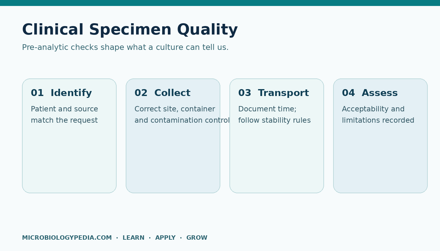
Before I interpret a smear, I check the specimen type, collection site, collection time, transport conditions, and any note that processing was delayed. A poorly collected or transported sample can make a technically correct stain less useful. The appearance of host cells and the mix of organisms may help assess some specimen types, but the criteria are specimen specific. I use the laboratory’s acceptance and reporting rules, not a universal shortcut.
Describe what the Gram stain actually shows
I record the Gram reaction, shape, and arrangement in plain language: for example, purple cocci in clusters or pink rods. I then check the stain controls and slide quality. A smear that is too thick, unevenly decolorized, or prepared from old growth can mislead. Gram-positive and Gram-negative are staining patterns; they are not species names, and an apparent match is only a starting hypothesis.
Bring culture into the picture
Next I ask whether growth is pure or mixed, how many distinct colony types appear, and whether the growth fits the specimen and smear. Colony size, pigment, hemolysis, and other features can guide the next validated identification steps. If the Gram stain and culture do not agree, I revisit sampling, contamination, and method performance before forcing an answer. A biochemical or molecular result also has to fit the sample context and its controls.
What I report and what I hold back
I distinguish a preliminary observation from a confirmed identification. I do not infer infection from one colony or turn a morphology clue into a treatment recommendation. In a clinical setting, significance depends on the specimen, patient context, organism, quantity, and local reporting policy. The laboratory’s approved workflow and qualified clinical interpretation govern the final result.
My quick review: Was this the right specimen? Are the controls valid? Does the smear fit the culture? What can the method actually identify? What uncertainty needs to appear in the report? That sequence is more reliable than jumping from a stain color straight to a species name.
Further reading
CDC Laboratory Training: Biochemicals and Gram-Positive Organism ID covers Gram-stain morphology, colony characteristics, and identification algorithms. This article is educational and does not replace a clinical laboratory procedure.

Noman Iftikhar is a microbiologist with an MPhil in Microbiology and professional experience in pharmaceutical microbiology. His work includes sterility testing, bacterial endotoxin testing, microbial limit testing, environmental monitoring, and microbiological quality control. Through MicrobiologyPedia, he shares practical, evidence-based guidance to help microbiology students and laboratory professionals understand testing procedures, laboratory practices, and quality standards.