
A simple stain colours bacterial cells with a single basic dye, such as methylene blue, crystal violet, safranin or carbol fuchsin. Every cell takes up the same colour, so the stain cannot separate groups of bacteria; what it shows clearly is cell shape, size and arrangement. This guide covers the principle, the procedure step by step, what to record as observations and how to interpret them.
Principle of simple staining
Bacterial cells carry a net negative charge at the near-neutral pH of most staining solutions, mainly from nucleic acids, teichoic acids and lipopolysaccharide. Basic dyes have a positively charged coloured ion (chromophore), so it binds to the negatively charged cell and colours it. The glass slide and background stay largely colourless, giving coloured cells against a clear field.
Acidic dyes such as nigrosin work the opposite way: their negatively charged chromophore is repelled by the cell and colours the background instead. That is the basis of negative staining, a different technique.
Stains used and typical staining times
| Basic dye | Colour of cells | Typical staining time |
|---|---|---|
| Methylene blue | Blue | 1–2 minutes |
| Crystal violet | Purple | 20–60 seconds |
| Safranin | Pink to red | 30–60 seconds |
| Carbol fuchsin | Red to magenta | 15–30 seconds |
Simple staining procedure step by step
- Prepare a clean slide. Use a grease-free glass slide and label it.
- Make a thin smear. From broth, spread one or two loopfuls. From a plate, mix a very small amount of growth into a drop of water. A thin, even film is essential.
- Air dry. Let the smear dry completely; it turns slightly cloudy.
- Heat fix. Pass the slide, smear side up, through a flame two or three times. Fixing kills the cells and attaches them to the slide. Overheating distorts cell shape.
- Apply the stain. Flood the smear with one basic dye for the time shown in the table above.
- Rinse. Wash off the excess dye gently with water.
- Blot dry. Press with absorbent paper; do not rub.
- Examine. Start with the low-power objective, then add immersion oil and examine with the 100× oil-immersion objective.
Simple stain observations
Record what is seen under oil immersion. A simple stain gives four kinds of information:
| Feature | What to record | Examples |
|---|---|---|
| Colour | All cells the colour of the dye used | Blue with methylene blue, purple with crystal violet |
| Shape | Cocci (spherical), bacilli (rods), coccobacilli, spirilla | Cocci; rods with rounded ends |
| Arrangement | Pairs, chains, clusters, groups of four, single cells | Diplococci, streptococci, staphylococci, tetrads, streptobacilli |
| Size and other features | Relative length and width; any granules or uneven staining | Long thin rods; beaded rods with dark granules |
Interpretation of simple staining results
- Shape and arrangement are the main results. For example, purple cocci in clusters suggest a staphylococcal type of arrangement, and cocci in chains a streptococcal type. These are clues for the next tests, not an identification.
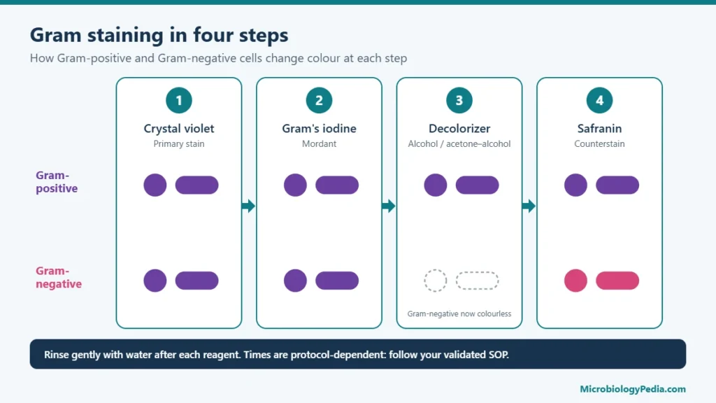
- All cells the same colour is the expected result. A simple stain cannot show whether an organism is Gram-positive or Gram-negative; that needs the Gram stain.
- Uneven or beaded staining in rods can indicate storage granules. Metachromatic (volutin) granules, which appear reddish-purple with methylene blue, are a classic feature of Corynebacterium.
- Mixed shapes on one slide suggest a mixed culture or contamination; subculture to obtain a pure culture before further work.
Example of a written result: “Simple stain with methylene blue: blue, uniformly stained cocci, approximately 1 µm, arranged in grape-like clusters.”
Common errors and how to avoid them
| Problem | Likely cause | Fix |
|---|---|---|
| Cells piled up, hard to see shape | Smear too thick | Use less material; spread a thinner film |
| Few or no cells | Smear too thin, or washed off | Use more material; air dry and heat fix fully before staining |
| Shrunken or distorted cells | Overheating during fixation | Pass the slide quickly through the flame two or three times only |
| Stain deposits or crystals | Old or unfiltered stain, or stain allowed to dry on the slide | Filter the stain; do not let it dry; rinse promptly |
| Pale cells | Staining time too short or dye too dilute | Extend the time within the procedure limits |
| Very dark, cells not separated | Overstaining or thick smear | Shorten the time; thinner smear |
Simple stain vs Gram stain vs negative stain
| Simple stain | Gram stain | Negative stain | |
|---|---|---|---|
| Dyes | One basic dye | Crystal violet, iodine, decolouriser, safranin | One acidic dye (for example nigrosin) |
| Heat fixed | Yes | Yes | No |
| What is coloured | The cells | Cells, by cell wall type | The background |
| Main use | Shape, size, arrangement | Separates Gram-positive and Gram-negative bacteria | True cell size and shape, capsules |
| Type | Simple | Differential | Simple (indirect) |
Related guides: Gram stain results of common bacteria, Gram-positive vs Gram-negative bacteria and from Gram stain to culture.
Frequently Asked Questions (FAQs)
What is the purpose of a simple stain?
To make bacterial cells visible so that their shape, size and arrangement can be seen under the microscope. It is quick and uses a single dye.
Why are basic dyes used in simple staining?
Bacterial cells are negatively charged, and basic dyes carry a positively charged chromophore that binds to them. The cells take up the colour while the background stays clear.
Can a simple stain tell Gram-positive from Gram-negative bacteria?
No. Every cell takes up the same dye. A differential stain, such as the Gram stain, is needed to separate the two groups.
Why is the smear heat fixed?
Heat fixing kills the cells and attaches them to the slide so they are not washed off during staining. Too much heat distorts their shape.
Which dye is best for a simple stain?
Methylene blue is the most common choice because it is gentle and seldom overstains. Crystal violet and safranin give stronger colour in a shorter time.
What magnification is used to read a simple stain?
The 100× oil-immersion objective, giving about 1,000× total magnification, after locating a thin area of the smear at low power.
References
- Cappuccino JG, Welsh CT. Microbiology: A Laboratory Manual. Pearson.
- Leboffe MJ, Pierce BE. A Photographic Atlas for the Microbiology Laboratory. Morton Publishing.
- Tille PM. Bailey & Scott’s Diagnostic Microbiology. Elsevier.
- Austin Community College. Stain protocols (simple, Gram and capsule stains). austincc.edu
- University of Wisconsin–Madison, Department of Bacteriology. Simple staining procedure. instr.bact.wisc.edu

Noman Iftikhar is a microbiologist with an MPhil in Microbiology and professional experience in pharmaceutical microbiology. His work includes sterility testing, bacterial endotoxin testing, microbial limit testing, environmental monitoring, and microbiological quality control. Through MicrobiologyPedia, he shares practical, evidence-based guidance to help microbiology students and laboratory professionals understand testing procedures, laboratory practices, and quality standards.