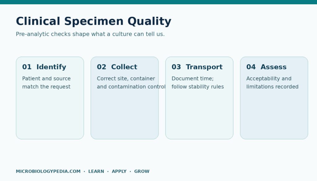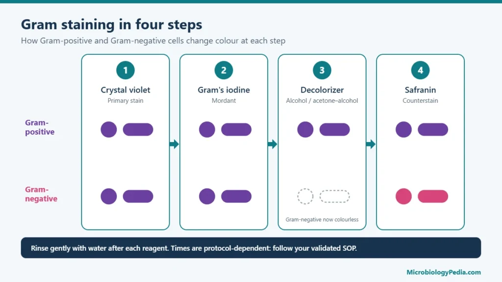
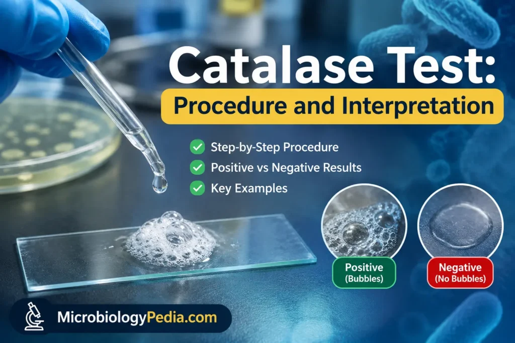
The catalase test detects catalase, an enzyme that breaks hydrogen peroxide (H2O2) down into water and oxygen. Growth from a pure, 18–24-hour colony is exposed to 3% H2O2. Immediate bubbling (released O2) is a positive catalase test; no bubbles is a negative catalase test. The test takes seconds, and its main use is to separate staphylococci (positive) from streptococci and enterococci (negative) after a Gram stain has shown Gram-positive cocci.
Strong reactions are easy to read. Weak ones need judgement, because a trace of blood agar, a metal loop, an old culture or degraded reagent can each distort the result. This guide covers the catalase test procedure, how to interpret strong, weak and doubtful reactions, and the controls that keep the result reliable.
What is the catalase test?
The catalase test is a rapid biochemical test used early in bacterial identification. It shows whether an isolate decomposes hydrogen peroxide:
2 H2O2 → 2 H2O + O2
The oxygen released forms the visible bubbles. The result is always read together with the Gram stain, cell morphology and colony appearance. A catalase result narrows the possibilities to a group of genera; it does not identify a species.
Catalase test principle
Bacteria that respire with oxygen produce reactive by-products, including superoxide and hydrogen peroxide. Hydrogen peroxide damages DNA, proteins and membranes, so organisms that live with oxygen need a way to remove it. Catalase is one of the main defences.
Most aerobic and facultatively anaerobic bacteria that contain cytochromes produce a haem-containing catalase. Streptococcus and Enterococcus are generally catalase-negative in conventional testing. However, some enterococci can produce weak reactions through pseudocatalase activity. Enterococcus faecalis also possesses a haem-dependent catalase system that can become active when haem is available. Therefore, these organisms should not be treated as metabolically identical. Many strict anaerobes also lack catalase, although there are exceptions.
In the test, the growth is exposed to hydrogen peroxide far in excess of anything the cell meets naturally. If catalase is present, oxygen is released faster than it can dissolve and escapes as bubbles.
Role of the catalase test in bacterial identification
After the Gram stain, catalase is usually the first branch for Gram-positive organisms: it separates staphylococci and micrococci from streptococci and enterococci, and Bacillus, Listeria and Corynebacterium from Lactobacillus and Erysipelothrix. It also helps separate aerotolerant Clostridium (negative) from Bacillus (positive). Most Gram-negative aerobes, including Enterobacterales, Pseudomonas and Neisseria, are positive, so the routine test adds less there; the superoxol test uses a higher hydrogen peroxide concentration, commonly 30%, for presumptive differentiation of certain Neisseria species, particularly N. gonorrhoeae. It is a specialised variation, not the routine catalase test.
Reagents and materials
| Item | Specification and notes |
|---|---|
| Hydrogen peroxide | 3% H2O2 for routine testing of aerobic and facultative bacteria (UK SMI TP 8 allows 3–6%). Store at 2–8 °C, protected from light; follow the manufacturer’s expiry and the laboratory’s QC frequency. |
| Culture | A well-isolated, 18–24-hour colony, preferably from a medium without whole blood, or a heavily inoculated 18–24-hour nutrient agar slant. |
| Transfer tool | Sterile wooden applicator stick, disposable plastic loop or glass rod, as specified in the laboratory’s validated procedure (see the metal loops section below). |
| Slide method | Clean glass slide inside a Petri dish with a lid. |
| Tube method | Clean 12 × 75 mm test tube or capped bijou bottle. |
| Reading aids | Dark background, magnifying lens; a microscope for very weak reactions. |
| Controls | A catalase-positive and a catalase-negative strain (see quality control). |
Higher concentrations belong to specific methods only: 15% H2O2 in some anaerobe schemes and 30% H2O2 (superoxol) for presumptive differentiation of certain Neisseria species, not for the routine catalase test. These are corrosive and need extra skin and eye protection.
Catalase test procedure (slide method, step by step)
The steps below follow the ASM slide protocol. Test the positive and negative controls the same way, at the same time, with the same reagent and the same type of transfer tool.
- Check the culture. Use a pure, well-isolated colony, 18–24 hours old, with a known Gram reaction.
- Prepare the slide. Place a clean glass slide inside a Petri dish; the lid will contain aerosols from bursting bubbles.
- Pick the growth. With a sterile wooden stick or disposable plastic loop, take growth from the top of the colony without touching the agar.
- Place the growth on the slide as a small, visible smear.
- Add the reagent. Place one drop of 3% H2O2 onto the growth. Do not mix.
- Close the dish immediately and watch the drop against a dark background.
- Read at once. Read the reaction immediately after adding hydrogen peroxide. Immediate, vigorous bubbling indicates a positive result. No bubbling indicates a negative result when the controls perform as expected. Weak or doubtful reactions should be repeated and interpreted according to the laboratory’s validated procedure.
- Discard and record. Dispose of the slide, stick and pipette as contaminated waste; record the test and control results.
Slide vs tube catalase test
All variations use the same reagent and the same interpretation. They differ in how well they show weak reactions and how well they contain aerosols.
Tube (bottle) method
- Add 4–5 drops of 3% H2O2 to a clean 12 × 75 mm tube.
- Either place the tip of a wooden stick carrying the growth into the peroxide, or rub the growth on the inner wall just above the liquid, cap the tube and tilt it so the peroxide covers the growth (UK SMI TP 8).
- Hold the tube against a dark background and look for bubbles rising from the growth.
Agar slant method
Add 1.0 mL of 3% H2O2 directly onto a heavily inoculated 18–24-hour pure culture on a nutrient agar slant, replace the cap and look for immediate bubbling. UK SMI TP 8 notes that many laboratories prefer this approach because the closed tube limits aerosols.
| Feature | Slide method | Tube or slant method |
|---|---|---|
| Growth needed | Very little | A little more; the slant needs a separate culture |
| Weak reactions | Small bubbles easy to see with a lens or microscope | Very weak reactions can be missed |
| Aerosol containment | Poor on an open slide; better inside a closed Petri dish | Better, especially with a capped tube or slant |
| Main risks | Agar carry-over, mixing the drop, drying | Dirty glassware |
Catalase test interpretation: positive, negative and weak results
| Observation | Interpretation | Action |
|---|---|---|
| Immediate, vigorous bubbling | Positive catalase test | Record as positive; continue the identification scheme (for staphylococci, usually a coagulase test). |
| One or a few bubbles appearing immediately, or seen only with a lens or microscope | Weak positive (to be confirmed) | Repeat as described below before reporting; read against the controls. |
| No bubbles | Negative catalase test | Record as negative if the positive control bubbled and the negative control did not. |
| A few tiny bubbles forming only after the reading time | Not a positive result | Record as negative; late bubbles are not interpreted. |
| Controls do not give the expected results | Invalid run | Check the reagent, tools and glassware, then repeat; do not report test results. |
Positive catalase test
A true positive is immediate and obvious: froth forms over the growth within a second or two, as with Staphylococcus aureus. The positive control should look the same.
Negative catalase test
The drop stays clear, with no bubbles rising from the growth during the reading time, as with Streptococcus pyogenes. A negative result is only valid if the positive control bubbled in the same run.
Weak positive, pseudocatalase and false-positive reactions
One or two bubbles should not be dismissed automatically. The CDC describes a weak positive as only one or two bubbles, and the ASM protocol recommends a lens or microscope to see them. Three different things can produce a small reaction, and they mean different things:
| Type | What causes it | Typical situations |
|---|---|---|
| Weak true catalase activity | Real catalase, but low activity: a small inoculum, an older culture, or an organism with naturally weak or variable activity | Aerococcus (may be weakly positive), Rothia (variable). Enterococcus faecalis: documented haem-dependent catalase activity when haem is available. Other lactic acid bacteria may produce manganese-dependent pseudocatalase, depending on the species and strain. |
| Pseudocatalase | A non-haem, manganese-containing enzyme that also decomposes H2O2; it does not need haem in the medium | Some lactic acid bacteria (for example strains of Lactobacillus plantarum and pediococci); some Enterococcus strains |
| False positive (artefact) | Peroxide decomposed by something other than the organism | Red blood cells carried over from blood agar, a reactive metal loop, dirty glassware |
These cannot be told apart at the bench from a single test. When to repeat: repeat whenever the reaction is weak, the colony came from a blood-containing medium, or the result conflicts with the Gram stain and colony appearance (for example, a “positive” coccus in chains).
- Subculture the isolate to a medium without blood or added haem (for example tryptic soy or nutrient agar) and incubate for 18–24 hours.
- Repeat the test with a visible amount of growth, fresh reagent, a non-metal tool, and positive and negative controls handled the same way.
- Read immediately with a lens. Report according to the laboratory’s procedure (positive, weak positive or negative), and confirm the identification with genus-appropriate tests, such as coagulase for staphylococci or PYR, bile–aesculin and 6.5% NaCl for enterococci.
Catalase-positive and catalase-negative bacteria
| Group | Catalase-positive | Catalase-negative |
|---|---|---|
| Gram-positive cocci | Staphylococcus (e.g. S. aureus, S. epidermidis), Micrococcus; Rothia variable; Aerococcus: variable catalase reactions; A. urinae is generally positive, whereas A. viridans is generally negative. Weak reactions may occur in some strains. | Streptococcus (e.g. S. pyogenes), Enterococcus, Abiotrophia, Gemella, Leuconostoc, Pediococcus |
| Gram-positive rods | Bacillus, Listeria, most Corynebacterium, Nocardia | Lactobacillus (some strains show pseudocatalase), Erysipelothrix, Arcanobacterium, most Clostridium |
| Gram-negative bacteria | Most Enterobacterales (e.g. Escherichia coli, Klebsiella), Pseudomonas aeruginosa, Neisseria | Less common among routine aerobic isolates; use genus-specific identification schemes |
Exceptions exist within otherwise positive genera: Staphylococcus aureus subsp. anaerobius and Staphylococcus saccharolyticus are catalase-negative, and rare catalase-negative strains of usually positive species have been reported. Gram reactions for many of these organisms are summarised in Gram stain results of common bacteria.
Staphylococcus vs Streptococcus: catalase test for Gram-positive cocci
Staphylococci and streptococci can look alike on a plate, and Gram stains made from colonies do not always show clear clusters or chains. The catalase test separates them in seconds: S. aureus and coagulase-negative staphylococci bubble vigorously, while S. pyogenes, S. agalactiae, viridans streptococci and S. pneumoniae do not.
- Catalase-positive cocci in clusters: continue with a coagulase test to separate S. aureus from coagulase-negative staphylococci. Micrococcus is also catalase-positive, so further tests are needed before reporting a staphylococcus.
- Catalase-negative cocci in pairs or chains: continue with haemolysis, PYR, bile–aesculin, 6.5% NaCl tolerance, optochin or Lancefield grouping, according to the organism suspected.
Enterococcus: pseudocatalase or haem-dependent catalase?
Enterococcus species are classed as catalase-negative, but some strains react weakly, and two different mechanisms have been described:
- Haem-dependent catalase. E. faecalis carries a gene for a true haem catalase (KatA) but cannot synthesise haem. Catalase activity was detected only when cells were grown with haem (hemin) added to the medium (Frankenberg et al., 2002).
- Pseudocatalase. Older work found catalase-positive E. faecalis (then Streptococcus faecalis) strains that could not make a haem-type enzyme (Johnston and Delwiche, 1965), consistent with the haem-independent pseudocatalase that UK SMI TP 8 notes in some Enterococcus strains.
Whether haem from blood agar is enough to switch on KatA in a routine culture has not been established in the sources cited here, and blood carry-over can itself cause bubbling. The practical rule is therefore the same for every weak reaction from a Gram-positive coccus in pairs or short chains: do not read it as “staphylococcus”. Repeat from a medium without blood or haem and confirm with enterococcal tests.
Common errors: false-positive and false-negative results
| Problem | Likely cause | Prevention |
|---|---|---|
| False positive from blood agar | Red blood cells contain catalase; a trace of blood picked up with the colony bubbles | Prefer colonies from a medium without whole blood. If blood agar is the only source, take growth from the top of the colony and confirm weak reactions by subculture. UK SMI TP 8 allows colonies from chocolate agar, where the red cells are lysed. |
| False positive from tools | Some metal loops decompose peroxide (see below) | Use the non-metal tool specified in the validated procedure. |
| False positive in tubes | Dirty glass tubes or bottles | Use clean or single-use tubes; run a negative control. |
| False negative from old cultures | Cultures older than 18–24 hours may lose catalase activity | Use 18–24-hour growth. |
| False negative from weak reagent | H2O2 has decomposed through warmth, light or age | Store cold and dark; check with controls daily or before use. |
| False negative from too little growth | Inoculum too small to show bubbles | Take a visible amount of growth; use a lens. |
| False negative with anaerobes | Tested straight from anaerobic incubation | Expose the culture to air for 30 minutes before testing (UK SMI TP 8). |
Metal loops and non-enzymatic peroxide decomposition
Hydrogen peroxide can be decomposed by some metal surfaces without any enzyme present, producing bubbles that look like a positive. Published methods disagree on which wire is the problem. The ASM protocol and one reagent manufacturer state that a platinum loop can cause a false positive and that nichrome does not, while UK SMI TP 8 states that some nichrome loops can react. Because the risk depends on the wire, its condition and the order of addition, the safest approach is to avoid metal altogether:
- Use a sterile wooden stick, disposable plastic loop or glass rod, as the CDC procedure recommends.
- If a validated procedure specifies a metal loop, place the growth on the slide first and add the peroxide afterwards; never touch the loop to the reagent.
- Handle the negative control with the same type of tool, so that any tool-related bubbling shows up in the control.
Quality control organisms and controls
Test a positive and a negative control in parallel at the frequency set by the laboratory’s procedure; UK SMI TP 8 recommends checking the peroxide daily or immediately before use. Grow the controls on the same type of medium as the test isolates.
| Control | Strain | Source | Expected result |
|---|---|---|---|
| Positive | Staphylococcus aureus ATCC 25923 | Reagent manufacturer’s instructions (Dalynn RC35) | Immediate, vigorous bubbling |
| Positive | Staphylococcus aureus NCTC 6571 or NCTC 12973 | UK SMI TP 8 | Immediate, vigorous bubbling |
| Negative | Streptococcus pyogenes ATCC 19615 | Reagent manufacturer’s instructions (Dalynn RC35) | No bubbles |
| Negative | Streptococcus mitis NCTC 10712 | UK SMI TP 8 | No bubbles |
Some laboratory procedures use Enterococcus faecalis ATCC 29212 as the negative control. Because enterococci can give weak reactions, a streptococcus is the less ambiguous choice. Handle control strains as reference cultures; the growth promotion test guide covers passage limits and strain handling.
Safety and containment
- Aerosols. Oxygen released in the test produces aerosols that can carry viable bacteria. UK SMI TP 8 requires work likely to generate aerosols to be done in a microbiological safety cabinet; the ASM protocol recommends a biosafety cabinet where possible and a closed Petri dish for the slide method. Capped tubes and slants reduce exposure further. Because catalase reactions can generate bacteria-containing aerosols, perform aerosol-generating procedures in a certified microbiological safety cabinet according to the applicable risk assessment and laboratory biosafety procedures. A covered Petri dish alone should not replace required containment.
- Risk assessment. The containment level depends on the organisms likely to be present and on the local risk assessment. When a hazard group 3 organism (such as Brucella or Bacillus anthracis) cannot be excluded, follow the laboratory’s rule-out and referral procedures rather than performing routine open-bench tests.
- Mycobacteria. The ASM protocol advises against using this procedure for Mycobacterium; specific methods apply.
- Reagent. Hydrogen peroxide is an irritant at 3% and corrosive at higher concentrations. Wear gloves and eye protection and follow the safety data sheet.
Catalase test in pharmaceutical microbiology (USP <1113>)
USP general chapter <1113> Microbial Characterization, Identification, and Strain Typing is an informational chapter. It names the catalase test, with the oxidase and coagulase tests, as a key biochemical screening test, and describes it as separating staphylococci (catalase-positive) from streptococci (catalase-negative). It does not prescribe a catalase procedure, reagent strength or control strains; those come from the laboratory’s validated SOP.
The chapter separates three levels of work, and the catalase test belongs to the first two:
- Screening and characterisation: colony and cell morphology, Gram reaction and key biochemical reactions, used for trending and many risk assessments, for example routine environmental monitoring.
- Identification to genus, species or strain: needed when a deeper assessment is required, such as sterility test positives, recoveries above recommended levels, and the confirmatory identification required by USP <62> (see the microbial limit test guide).
Applications and limitations
Applications: first-line separation of Gram-positive cocci and rods, and screening isolates before choosing an identification kit or card.
Limitations: the catalase test is presumptive and never identifies a species on its own. Weak and atypical reactions occur (Enterococcus, Aerococcus, Rothia, pseudocatalase-producing lactic acid bacteria), catalase-negative variants of positive species exist, and results depend on medium, culture age, reagent strength and technique. A mixed culture gives a meaningless result.
Frequently asked questions
What does a positive catalase test mean?
Immediate bubbling shows that the organism decomposes hydrogen peroxide, normally because it makes catalase. Among Gram-positive cocci, a positive result points to Staphylococcus or Micrococcus rather than Streptococcus or Enterococcus.
What does a negative catalase test mean?
No bubbling on immediate reading, with controls performing as expected, means no detectable catalase. Among Gram-positive cocci this points to streptococci, enterococci or related genera.
What concentration of hydrogen peroxide is used?
3% H2O2 is the routine reagent. 15% is used in some anaerobe schemes; 30% is used in the superoxol test, a specialised variation for presumptive differentiation of certain Neisseria species, not the routine catalase test.
Can a catalase test be done from blood agar?
It is best avoided, because red blood cells contain catalase and carry-over can cause a false positive. If blood agar is the only source, take growth from the top of the colony and confirm any weak reaction from a non-blood medium. Chocolate agar is acceptable according to UK SMI TP 8.
Is Staphylococcus aureus catalase-positive?
Yes, routinely isolated S. aureus gives a strong positive and is the usual positive control. S. aureus subsp. anaerobius is an exception, and coagulase-negative staphylococci are also positive, so a coagulase test follows.
Is Streptococcus catalase-negative?
Yes. Streptococci, including S. pyogenes and S. pneumoniae, are catalase-negative; S. pyogenes and S. mitis are used as negative controls.
Is Enterococcus catalase-positive or negative?
Enterococci are classed as catalase-negative. Some strains give weak reactions, from a haem-dependent catalase (E. faecalis with haem in the medium) or a pseudocatalase, which must not be mistaken for a staphylococcal positive.
What is pseudocatalase?
Pseudocatalase is a non-haem, manganese-containing enzyme that decomposes hydrogen peroxide. It occurs in some lactic acid bacteria, such as strains of Lactobacillus plantarum, and can give a weak reaction in organisms otherwise regarded as catalase-negative.
Is E. coli catalase-positive?
Yes. Escherichia coli and most other Enterobacterales are catalase-positive, so the test is of limited value for separating them from each other.
What is the difference between the catalase and coagulase tests?
The catalase test separates staphylococci (positive) from streptococci (negative). The coagulase test is then performed on catalase-positive staphylococci to separate S. aureus (coagulase-positive) from coagulase-negative staphylococci.
Key points
The catalase test procedure is short: an 18–24-hour pure colony from a medium without whole blood, a non-metal tool, one drop of 3% H2O2 and an immediate read against controls. Interpretation needs more care. Strong, immediate bubbling is positive; no bubbling is negative when the controls perform as expected; anything weak is repeated before it is reported. Used this way, the catalase test remains one of the fastest first steps in identifying Gram-positive bacteria, ahead of tests such as coagulase or PYR. For the steps before it, see from Gram stain to culture and Gram-positive vs Gram-negative bacteria.
References
- UK Health Security Agency. UK Standards for Microbiology Investigations TP 8: Catalase test. Issue 4.1, 18 February 2025.
- UK Health Security Agency. UK Standards for Microbiology Investigations ID 1: Introduction to the identification of medically important bacteria and fungi from culture. Issue 4, 6 August 2025.
- Reiner K. Catalase Test Protocol. American Society for Microbiology; 11 November 2010.
- Centers for Disease Control and Prevention. Core Microbiology Skills: How to Perform a Catalase Test (video job aid transcript). CDC REACH.
- Frankenberg L, Brugna M, Hederstedt L. Enterococcus faecalis heme-dependent catalase. J Bacteriol. 2002;184(22):6351–6356. doi:10.1128/JB.184.22.6351-6356.2002
- Johnston MA, Delwiche EA. Distribution and characteristics of the catalases of Lactobacillaceae. J Bacteriol. 1965;90(2):347–351. doi:10.1128/jb.90.2.347-351.1965
- Kono Y, Fridovich I. Isolation and characterization of the pseudocatalase of Lactobacillus plantarum. J Biol Chem. 1983;258(10):6015–6019.
- Taylor WI, Achanzar D. Catalase test as an aid to the identification of Enterobacteriaceae. Appl Microbiol. 1972;24(1):58–61. doi:10.1128/am.24.1.58-61.1972
- Dalynn Biologicals. Catalase Reagent, Catalogue No. RC35 (instructions for use, revised January 2014).
- United States Pharmacopeia. General chapter <1113> Microbial Characterization, Identification, and Strain Typing. USP–NF.
This guide is for education. Laboratories should follow their validated procedures, the reagent manufacturer’s instructions and the current editions of the referenced standards.

Noman Iftikhar is a microbiologist with an MPhil in Microbiology and professional experience in pharmaceutical microbiology. His work includes sterility testing, bacterial endotoxin testing, microbial limit testing, environmental monitoring, and microbiological quality control. Through MicrobiologyPedia, he shares practical, evidence-based guidance to help microbiology students and laboratory professionals understand testing procedures, laboratory practices, and quality standards.