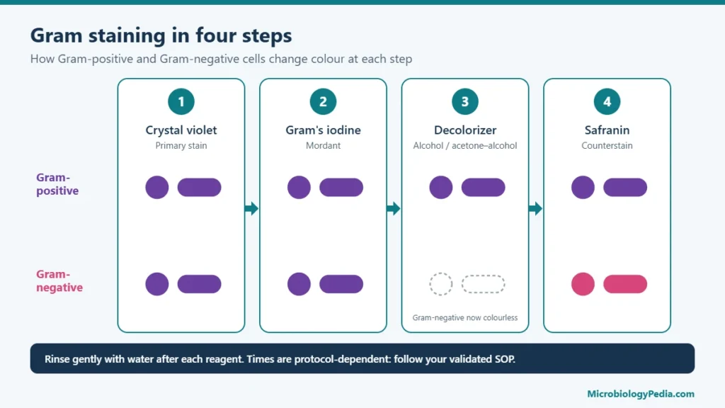
Gram-positive bacteria have a thick peptidoglycan cell wall and no outer membrane, so they keep the crystal violet dye and stain purple. Gram-negative bacteria have a thin peptidoglycan layer covered by an outer membrane that contains lipopolysaccharide (LPS, endotoxin); they lose the crystal violet during decolorization and stain pink with the counterstain. That structural difference explains most of the practical differences between the two groups: staining, endotoxin, antibiotic and disinfectant susceptibility, and where each group turns up in a laboratory or a manufacturing facility.
Gram-positive vs Gram-negative bacteria: comparison table
| Feature | Gram-positive | Gram-negative |
|---|---|---|
| Gram stain result | Purple (retain crystal violet) | Pink to red (take up safranin) |
| Peptidoglycan | Thick, 30–100 nm, many layers | Thin, a few nanometres, one to a few layers |
| Outer membrane | Absent | Present |
| Lipopolysaccharide (endotoxin) | Absent | Present in the outer membrane |
| Teichoic and lipoteichoic acids | Present | Absent |
| Periplasm | No defined compartment between two membranes | Present, between inner and outer membranes |
| Porins | Absent | Present in the outer membrane |
| Endospore formation | Some genera (Bacillus, Clostridium) | Not a feature of the common Gram-negative genera |
| Typical examples | Staphylococcus, Streptococcus, Bacillus, Clostridium, Listeria | Escherichia, Salmonella, Klebsiella, Pseudomonas, Neisseria |
Cell wall structure: the reason for the difference
Gram-positive cell wall
The Gram-positive envelope has one membrane, the cytoplasmic membrane, surrounded by many layers of peptidoglycan that together are about 30–100 nm thick. Long anionic polymers called teichoic acids run through the peptidoglycan; wall teichoic acids are linked to the peptidoglycan and lipoteichoic acids are anchored in the membrane. There is no outer membrane, so the peptidoglycan is the outermost major layer apart from any capsule or surface proteins.
Gram-negative cell wall
The Gram-negative envelope has two membranes. The inner (cytoplasmic) membrane is followed by the periplasm, which contains a thin peptidoglycan layer only a few nanometres thick, and then by the outer membrane. The outer leaflet of the outer membrane is made largely of lipopolysaccharide. Its lipid A portion is the endotoxin that causes fever when it reaches the bloodstream. Small hydrophilic molecules cross the outer membrane through protein channels called porins; large or hydrophobic molecules are largely kept out.
Why they stain differently
In the Gram stain, crystal violet and iodine form a large complex inside all cells. The alcohol or acetone decolorizer dehydrates the thick peptidoglycan of Gram-positive cells, which traps the complex, so the cells stay purple. In Gram-negative cells the decolorizer disrupts the outer membrane and the thin peptidoglycan cannot hold the complex; the cells become colourless and then take up the pink safranin counterstain. The full method, including timing and controls, is in the Gram staining procedure.
Examples of Gram-positive bacteria
| Shape | Genera and species | Typical arrangement |
|---|---|---|
| Cocci | Staphylococcus aureus, S. epidermidis, Micrococcus luteus | Clusters (staphylococci), tetrads (micrococci) |
| Cocci | Streptococcus pyogenes, S. pneumoniae, Enterococcus faecalis | Chains or pairs |
| Bacilli, spore-forming | Bacillus subtilis, B. cereus, Clostridium sporogenes | Large rods, often in chains |
| Bacilli, non-spore-forming | Listeria monocytogenes, Corynebacterium, Lactobacillus, Cutibacterium acnes | Short or irregular rods |
Examples of Gram-negative bacteria
| Shape | Genera and species | Notes |
|---|---|---|
| Bacilli (rods), fermenters | Escherichia coli, Klebsiella, Salmonella, Shigella, Enterobacter, Proteus | Order Enterobacterales; oxidase negative |
| Bacilli (rods), non-fermenters | Pseudomonas aeruginosa, Burkholderia cepacia complex, Ralstonia pickettii, Acinetobacter | Common in water and wet environments |
| Cocci and diplococci | Neisseria meningitidis, N. gonorrhoeae, Moraxella catarrhalis | Kidney-shaped pairs |
| Coccobacilli | Haemophilus influenzae | Small, pleomorphic |
| Curved or spiral rods | Vibrio cholerae, Campylobacter jejuni, Helicobacter pylori | Comma or spiral shaped |
For the microscopic appearance of individual species, see Gram stain results of common bacteria.
Antibiotic and disinfectant susceptibility
The outer membrane is a permeability barrier, and it is the main reason some antibiotics act on only one group. Vancomycin, a large glycopeptide, cannot cross the outer membrane and is therefore used against Gram-positive bacteria. Polymyxins such as colistin bind lipopolysaccharide and act on Gram-negative bacteria. Many agents, including several beta-lactams, fluoroquinolones and aminoglycosides, cover members of both groups to different degrees.
Group membership does not predict the susceptibility of an individual isolate. Acquired resistance is common in both groups, and many Gram-negative bacteria are intrinsically resistant to several antibiotic classes, so the choice of an antibiotic rests on susceptibility testing and current clinical guidance, not on the Gram reaction alone.
The same barrier affects chemical disinfection. As a group, Gram-negative bacteria, notably Pseudomonas and Burkholderia, tend to be less susceptible to some disinfectants and preservatives than vegetative Gram-positive bacteria, while bacterial endospores from Gram-positive Bacillus and Clostridium are far more resistant than any vegetative cell and require a sporicidal agent.
Why the distinction matters in pharmaceutical microbiology
- Endotoxin. Only Gram-negative bacteria carry lipopolysaccharide. Gram-negative contamination of water, raw materials or equipment is the source of the endotoxin measured by the bacterial endotoxins test, and endotoxin remains after the cells are killed.
- Water systems. Isolates from purified water and wet areas are mostly Gram-negative rods such as Pseudomonas, Burkholderia cepacia complex and Ralstonia.
- Cleanroom monitoring. Gram-positive cocci from skin (Staphylococcus, Micrococcus) are the usual finding in environmental monitoring of occupied cleanrooms; Gram-positive spore-forming rods point to materials, cardboard, soil or dust; Gram-negative rods point to water or inadequate drying.
- Specified microorganisms. The tests in USP <62> cover both groups: Staphylococcus aureus and clostridia are Gram-positive; Escherichia coli, Salmonella, Pseudomonas aeruginosa and the bile-tolerant Gram-negative bacteria are Gram-negative. See the microbial limit test.
- Investigations. A Gram stain of an isolate is the first step in identification and directs the next tests: catalase and coagulase for Gram-positive cocci, oxidase and fermentation tests for Gram-negative rods.
Bacteria that do not fit either group cleanly
- Mycobacteria have a waxy, mycolic-acid-rich wall, stain poorly by Gram and are detected with an acid-fast stain.
- Mycoplasmas have no cell wall and do not retain the stain.
- Gram-variable results occur with old cultures of Gram-positive bacteria and with some genera, including Actinomyces, Arthrobacter and Corynebacterium.
- Acinetobacter is Gram-negative but can resist decolorization and appear Gram-positive.
Frequently Asked Questions (FAQs)
What is the main difference between Gram-positive and Gram-negative bacteria?
The cell envelope. Gram-positive bacteria have a thick peptidoglycan wall and no outer membrane; Gram-negative bacteria have a thin peptidoglycan layer and an outer membrane containing lipopolysaccharide.
What colour are Gram-positive and Gram-negative bacteria?
After a Gram stain, Gram-positive bacteria are purple and Gram-negative bacteria are pink to red.
Are Gram-negative bacteria more dangerous than Gram-positive bacteria?
Neither group is more dangerous as a whole; both include harmless organisms and serious pathogens. Gram-negative bacteria carry endotoxin and are often intrinsically resistant to more antibiotics, while Gram-positive pathogens such as Staphylococcus aureus and Streptococcus pneumoniae cause severe infections of their own.
Is E. coli Gram-positive or Gram-negative?
Escherichia coli is a Gram-negative rod.
Is Staphylococcus aureus Gram-positive or Gram-negative?
Staphylococcus aureus is a Gram-positive coccus that grows in clusters.
Do Gram-positive bacteria produce endotoxin?
No. Endotoxin is the lipopolysaccharide of the Gram-negative outer membrane. Gram-positive bacteria may release other substances, including exotoxins, but not endotoxin.
Which Gram-negative bacteria are cocci?
Neisseria species and Moraxella catarrhalis are Gram-negative cocci that usually appear as diplococci. Most other common Gram-negative bacteria are rods.
References
- Silhavy TJ, Kahne D, Walker S. The bacterial cell envelope. Cold Spring Harbor Perspectives in Biology. 2010;2(5):a000414. doi:10.1101/cshperspect.a000414
- Smith AC, Hussey MA. Gram Stain Protocols. American Society for Microbiology; 2005. asm.org/protocols/gram-stain-protocols
- Gram-Positive Bacteria. In: StatPearls [Internet]. Treasure Island (FL): StatPearls Publishing. NCBI Bookshelf NBK470553
- Oliveira J, Reygaert WC. Gram-Negative Bacteria. In: StatPearls [Internet]. Treasure Island (FL): StatPearls Publishing; updated 2023. PMID 30855801
- United States Pharmacopeia. General Chapter <62> Microbiological Examination of Nonsterile Products: Tests for Specified Microorganisms.
Related guides on MicrobiologyPedia
- Gram staining: principle, procedure and results
- Gram stain results of common bacteria
- Bacterial endotoxin and endotoxin testing
- Microbial limit test (USP 61 and 62)
- Environmental monitoring in pharmaceutical cleanrooms
After the Gram reaction, Gram-positive cocci are usually split with the catalase test: staphylococci are catalase-positive, while streptococci and enterococci are catalase-negative. See Catalase Test: Principle, Procedure and Interpretation of Results.

Noman Iftikhar is a microbiologist with an MPhil in Microbiology and professional experience in pharmaceutical microbiology. His work includes sterility testing, bacterial endotoxin testing, microbial limit testing, environmental monitoring, and microbiological quality control. Through MicrobiologyPedia, he shares practical, evidence-based guidance to help microbiology students and laboratory professionals understand testing procedures, laboratory practices, and quality standards.