Gram staining is a differential staining method that sorts most bacteria into two groups by how their cell envelope behaves during decolorization. Gram-positive bacteria keep the crystal violet–iodine complex and look purple. Gram-negative bacteria lose it and take up a red counterstain, so they look pink. A Gram stain also shows cell shape and arrangement, and it gives this information within minutes. That speed is why it remains one of the first tests in clinical, pharmaceutical and food microbiology laboratories.
This guide explains the principle, each reagent, the procedure, how to read the slide, and what goes wrong. It separates the established science from the simplified classroom version, and it treats reagent times as protocol-dependent. Your laboratory’s validated SOP and the reagent manufacturer’s instructions always take precedence.
What is Gram staining?
Gram staining is a differential stain: it uses more than one dye so that different kinds of cells end up different colours. The method is named after the Danish physician Hans Christian Gram, who published it in 1884 while looking for a way to make bacteria stand out from tissue. The modern four-reagent version developed through later modifications, including Hucker’s crystal violet–ammonium oxalate formulation of 1921.
More than a century later, the Gram stain is still the starting point for describing a bacterium. It answers three questions in one slide:
- Gram reaction: positive (purple) or negative (pink)?
- Shape: cocci, rods (bacilli), coccobacilli or curved forms?
- Arrangement: pairs, chains, clusters or single cells?
Those answers narrow the possibilities and decide which culture media, biochemical tests or identification systems come next. They do not identify a species on their own.
Principle of Gram staining
The Gram reaction reflects differences in the bacterial cell envelope, meaning everything outside the cytoplasm.
The Gram-positive cell envelope
A Gram-positive cell has a cytoplasmic membrane surrounded by a thick, multilayered peptidoglycan wall. Peptidoglycan is a mesh of sugar chains cross-linked by short peptides. Teichoic acids and lipoteichoic acids are threaded through it and give the wall a strong negative charge. There is no outer membrane.
The Gram-negative cell envelope
A Gram-negative cell has a cytoplasmic membrane and only a thin layer of peptidoglycan in the periplasmic space. Outside that is an outer membrane, a lipid bilayer whose outer leaflet contains lipopolysaccharide (LPS) and whose porin proteins let small molecules cross. Lipoproteins anchor the outer membrane to the peptidoglycan.

How the crystal violet–iodine complex forms
Crystal violet is a positively charged dye. It enters every cell on the slide and binds to negatively charged cell components, so at this stage all bacteria are purple.
Gram’s iodine is then added. Iodide combines with crystal violet to form an insoluble crystal violet–iodine (CV–I) complex. Chemical and electron-microscopy studies show this precipitate forms inside both Gram-positive and Gram-negative cells. It is much larger than the dye alone and harder to wash out.
What the decolorizer does to each cell type
The decolorizer (ethanol, or a mixture of acetone and alcohol) is the step that separates the two groups:
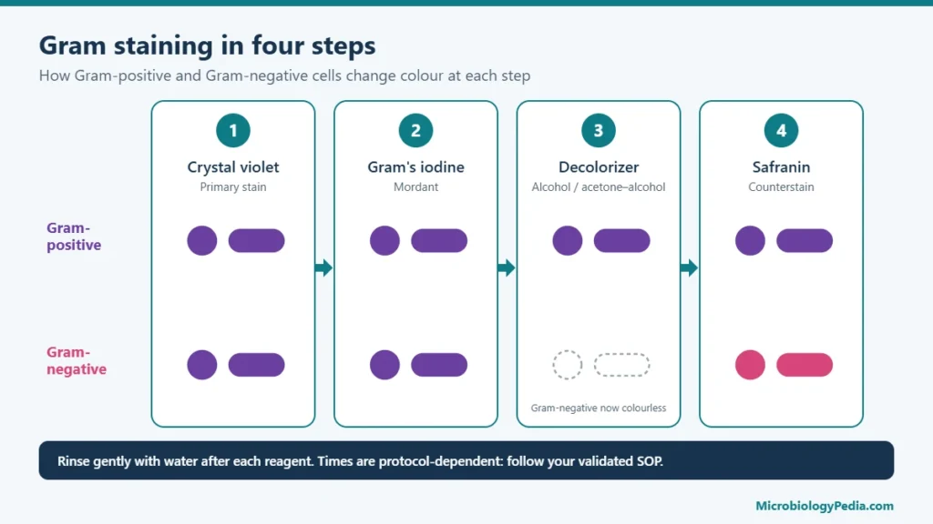
- In Gram-positive cells, the solvent dehydrates the thick peptidoglycan. The mesh tightens, and the CV–I complex stays trapped inside, so the cell stays purple.
- In Gram-negative cells, the solvent disrupts the lipid-rich outer membrane; in electron-microscopy studies of E. coli, the outer membrane was stripped away during ethanol decolorization. The thin peptidoglycan cannot hold the complex, so it washes out and the cell becomes colourless.
Safranin (or basic fuchsin) then stains the colourless Gram-negative cells pink or red. Gram-positive cells also take up the counterstain, but the dark purple hides it.

How Does Gram Staining Differentiate Gram-Positive and Gram-Negative Bacteria?
The Gram staining reaction depends on the structure and integrity of the bacterial cell envelope and its ability to retain the crystal violet–iodine (CV–I) complex during decolorization.
Gram-positive bacteria have a thick peptidoglycan layer that helps retain the crystal violet–iodine complex during alcohol or alcohol-acetone treatment. Consequently, these bacteria remain purple or violet.
Gram-negative bacteria have a thin peptidoglycan layer and an additional outer membrane. During decolorization, the cell envelope becomes more permeable, allowing the crystal violet–iodine complex to escape. These cells subsequently absorb safranin and appear pink or red.
Why Do Gram-Positive Bacteria Retain Crystal Violet?
The commonly accepted explanation involves changes in cell wall permeability during decolorization. Alcohol dehydrates the thick peptidoglycan layer, reducing its permeability and helping retain the crystal violet–iodine complex.
In contrast, alcohol disrupts the outer membrane of Gram-negative bacteria and facilitates the removal of the crystal violet–iodine complex.
This explanation is widely used to describe the differential staining reaction, although the exact molecular interactions responsible for dye retention are not completely understood.
How Does Cell Wall Damage Affect Gram Staining?
An intact bacterial cell wall is important for a reliable Gram staining reaction. Aging, autolysis, antibiotic exposure, and excessive heat can damage the cell envelope and affect its ability to retain crystal violet.
As a result, some Gram-positive bacteria may appear pink or show variable staining instead of their typical purple appearance.
Scientific note: The electron microscopy study by Beveridge and Davies (1983) provided important evidence regarding the localization of the crystal violet–iodine complex in bacterial cells. Their findings helped explain the differential staining reaction.explanation vs established evidenc
Reagents used in Gram staining
1. Crystal violet: the primary stain
Crystal violet stains every cell purple. If it is omitted, nothing is coloured until the counterstain, so every cell looks pink. If the smear is too thick, dye is trapped in clumps and Gram-negative cells can look purple.
2. Gram’s iodine: the mordant
Iodine fixes crystal violet inside the cell as the CV–I complex. If it is omitted or too weak, the dye washes out of Gram-positive cells too, giving false Gram-negative results. Unstabilized iodine solutions oxidize over time, which is why many commercial kits use stabilized forms such as polyvinylpyrrolidone–iodine.
3. Decolorizer: alcohol or acetone–alcohol
The decolorizer removes the complex from Gram-negative cells only. Its strength sets the timing: 95% ethanol acts slowest, acetone fastest, and acetone–alcohol mixtures fall between. This is the step that decides most errors:
- Too long, or too strong: Gram-positive cells lose colour and look Gram-negative.
- Too short: Gram-negative cells keep the purple and look Gram-positive.
4. Safranin: the counterstain
Safranin colours the decolorized Gram-negative cells pink to red. Some laboratories prefer basic fuchsin or carbol fuchsin, which stain weakly staining Gram-negative bacteria (for example Haemophilus and Legionella species, and some anaerobes) more intensely. If the counterstain is omitted, Gram-negative cells stay colourless and are easily missed.
| Reagent | Role | Gram-positive cells | Gram-negative cells | If omitted or misused |
|---|---|---|---|---|
| Crystal violet | Primary stain | Purple | Purple | All cells end up pink |
| Gram’s iodine | Mordant (forms CV–I complex) | Purple | Purple | Gram-positive cells decolorize (false negative) |
| Alcohol or acetone–alcohol | Decolorizer | Purple | Colourless | Over: Gram-positive cells look pink. Under: Gram-negative cells look purple |
| Safranin or basic fuchsin | Counterstain | Purple (counterstain masked) | Pink/red | Gram-negative cells stay colourless |
Gram stain procedure: step by step
Materials and quality control
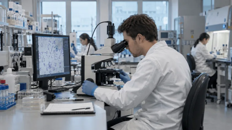
You need clean glass slides, an inoculating loop, a staining rack, a wash bottle or gentle tap water, the four reagents, a timer, and a brightfield microscope with a 100× oil immersion objective and immersion oil. Wear the PPE your laboratory’s risk assessment requires.
Run a control slide with each staining run or new reagent lot, as your procedure specifies. A typical control contains a Gram-positive coccus such as Staphylococcus aureus and a Gram-negative rod such as Escherichia coli. If the control does not give purple cocci and pink rods, the patient or sample slides cannot be trusted.
The steps
- Prepare a thin smear. For colonies, mix a small amount of growth into a drop of water or saline on the slide and spread it thinly. For a broth culture, spread a loopful directly.
- Air-dry the smear completely.
- Fix the smear. Pass it briefly through a flame or place it on a slide warmer, or flood it with methanol if your laboratory uses methanol fixation. Methanol is often preferred for direct clinical smears because it preserves cell structure. Do not overheat: excessive heat distorts cells and can make Gram-positive bacteria stain pink.
- Apply crystal violet to cover the smear.
- Rinse gently with water.
- Apply Gram’s iodine.
- Rinse gently with water.
- Decolorize. Hold the slide at an angle and apply the decolorizer drop by drop until the runoff is just clear. With acetone–alcohol this often takes only a few seconds.
- Rinse immediately with water to stop decolorization.
- Apply safranin (or basic fuchsin).
- Rinse, then air-dry or blot gently with bibulous paper. Do not wipe, because that removes cells.
- Examine under oil immersion with the 100× objective, after scanning first at lower power to find well-spread areas.
Typical reagent times and why they vary
Reagent times are protocol-dependent. The table shows the range you will meet. Use the times in your validated SOP or the manufacturer’s instructions.
| Step | Time for each Stain |
|---|---|
| Crystal violet | Up to 1 minute |
| Gram’s iodine | Up to 1 minute |
| Decolorizer (acetone–alcohol) | 15 Seconds; longer with 95% ethanol |
| Safranin or basic fuchsin | 30 seconds to 1 minute |
Four details that decide the result
- Smear thickness. You should see separate cells. Thick areas hold dye and give false Gram-positive results, so read thin areas only.
- Culture age. Stain young growth, typically 18–24 hours old. Older cultures contain cells with weakened walls that may stain pink.
- Decolorization. This is the most common source of error. Stop as soon as the runoff is clear and rinse at once.
- Washing. Rinse gently and briefly. A hard or long stream can wash cells off or pull stain from them.
How to interpret Gram staining results
Read only well-stained, thin areas, and check the control slide first. Then record the Gram reaction, the shape and the arrangement of each type of cell.
| Characteristic | Gram-positive | Gram-negative |
|---|---|---|
| Colour under the microscope | Purple to violet | Pink to red |
| Peptidoglycan layer | Thick, multilayered | Thin |
| Outer membrane | Absent | Present (contains LPS) |
| Teichoic acids | Present | Absent |
| Crystal violet–iodine complex after decolorization | Retained | Lost |
| Counterstain | Taken up but masked by purple | Visible as pink/red |
These differences go beyond colour. The outer membrane, for example, explains why many Gram-negative bacteria are intrinsically less susceptible to some antibiotics and detergents. Even so, the Gram reaction alone never tells you which antibiotic will work. Susceptibility needs its own testing.

Gram-positive vs Gram-negative bacteria: examples
| Organism | Gram reaction | Typical shape and arrangement |
|---|---|---|
| Staphylococcus aureus | Positive | Cocci in irregular clusters |
| Streptococcus pyogenes | Positive | Cocci in chains |
| Enterococcus faecalis | Positive | Cocci in pairs and short chains |
| Bacillus subtilis | Positive | Rods, often in chains; may show spores |
| Escherichia coli | Negative | Straight rods |
| Klebsiella pneumoniae | Negative | Plump rods, often with a capsule |
| Pseudomonas aeruginosa | Negative | Slender rods |
| Neisseria species | Negative | Diplococci with flattened sides, often called kidney-bean shaped |
Bacterial morphology and arrangement
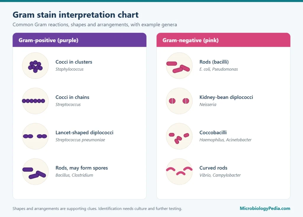
The Gram reaction, the cell shape and the arrangement are three separate observations. A Gram-positive coccus and a Gram-negative coccus share a shape but not a reaction, so record all three.
| Term | What you see | Example |
|---|---|---|
| Cocci | Spherical cells | Staphylococcus, Streptococcus |
| Bacilli (rods) | Rod-shaped cells | Bacillus, E. coli |
| Coccobacilli | Short, oval rods | Haemophilus, Acinetobacter |
| Diplococci | Cocci in pairs | Neisseria, Streptococcus pneumoniae |
| Streptococci (arrangement) | Cocci in chains | Streptococcus pyogenes |
| Staphylococci (arrangement) | Cocci in grape-like clusters | Staphylococcus aureus |
| Streptobacilli | Rods in chains | Bacillus species |
The arrangement depends partly on how a species divides: in one plane for chains, in several planes for clusters. Smear preparation can break chains and clusters apart, so arrangement is supporting evidence, not proof.
Factors that affect Gram staining results
- Over-decolorization removes the complex from Gram-positive cells, producing false Gram-negative results.
- Under-decolorization leaves Gram-negative cells purple, producing false Gram-positive results.
- Thick smears trap dye and prevent even decolorization.
- Old cultures contain cells with damaged walls that stain pink or unevenly.
- Incorrect fixation, such as overheating, distorts cells and damages walls. Under-fixed smears wash off.
- Poor reagents, such as precipitated crystal violet, oxidized iodine or expired stock, give weak staining or artifacts.
- Inadequate washing leaves reagent behind and continues decolorization. Washing too hard strips cells.
- Wrong reagent order, such as decolorizer before iodine, invalidates the stain.
- Antibiotic exposure before sampling can weaken walls and change the apparent Gram reaction.
Common Gram staining errors and troubleshooting
| Observation | Possible cause | Corrective action |
|---|---|---|
| Gram-positive bacteria appear pink | Over-decolorization; old culture; overheating during fixation; antibiotic-damaged cells | Time the decolorizer; check the control slide; stain an 18–24 h subculture; fix more gently |
| Gram-negative bacteria appear purple | Smear too thick; under-decolorization | Make a thinner smear; decolorize until the runoff is just clear; read thin areas |
| Mixed purple and pink cells of one morphology | Uneven smear thickness; aging culture; a genuinely Gram-variable organism | Re-stain fresh growth on a thin smear; if the pattern persists, consider a Gram-variable organism |
| Purple crystals or precipitate on the slide | Crystal violet precipitate; slide dried during staining; dirty slide | Filter crystal violet; keep the smear wet between steps; use clean slides |
| Cells hard to see, or none visible | Smear too thin or washed off; not fixed; wrong focal plane | Use more material; fix before staining; rinse gently; mark the smear area |
| Distorted or fragmented cells | Excessive heat fixation; very old culture; antibiotic effect | Fix gently or use methanol; subculture to fresh growth |
Gram-variable and atypical bacteria
Some bacteria do not give a clean Gram-positive or Gram-negative result. The reasons differ:
- Gram-positive organisms that lose stain as they grow and age. Genera such as Actinomyces, Corynebacterium, Bacillus and Clostridium can show a mixture of purple and pink cells. Electron microscopy found two routes. In Actinomyces, Corynebacterium and related genera, cells that had begun dividing were more fragile at the division site. In Bacillus, Butyrivibrio and Clostridium, the wall under the surface layer thinned as cultures aged, until stationary-phase cultures stained almost entirely Gram-negative.
- Gram-negative organisms that resist decolorization. Acinetobacter species are Gram-negative coccobacilli that often hold some crystal violet and can look Gram-positive.
- Mycobacteria. Mycobacterium species have a waxy, mycolic-acid-rich wall that resists the Gram reagents. They often stain weakly or not at all. Acid-fast staining (Ziehl–Neelsen or Kinyoun) or fluorescent auramine staining is used instead.
- Mycoplasma. Mycoplasma species have no cell wall, so they have no Gram reaction and are not reliably seen on a Gram stain. Culture, molecular methods or other stains are needed.
“Gram-variable” should describe an organism’s real behaviour, not a poorly made smear. Before calling an organism Gram-variable, repeat the stain on fresh growth with an acceptable control slide.
Applications of Gram staining
- Clinical microbiology. Direct smears from specimens such as cerebrospinal fluid, blood culture bottles and sputum give an early clue while culture is pending.
- Pharmaceutical microbiology. Gram staining is a routine first step in characterizing isolates from environmental monitoring, water systems, sterility test positives and media fills. The result guides the identification method chosen next, such as biochemical panels, MALDI-TOF mass spectrometry or sequencing. It also helps trend contamination patterns. USP general chapter <1113> covers this wider field of microbial characterization and identification.
- Food microbiology. Isolates from spoilage or hygiene investigations are often Gram-stained to steer confirmation tests.
- Environmental and research microbiology. Gram staining helps describe pure cultures. It reveals little about mixed natural communities, many of whose members cannot be cultured.
In each setting, Gram staining narrows the field. It does not establish species identity or antimicrobial susceptibility.
Limitations of Gram staining
- It cannot identify a species, and many unrelated genera look alike.
- It depends on technique: decolorization, smear thickness and culture age all change the result.
- Some organisms stain poorly or not at all, such as mycobacteria and mycoplasmas, or variably, such as aging Gram-positive genera.
- A direct specimen needs enough organisms to be seen. A negative smear does not rule out infection or contamination.
- It says nothing about viability, toxin production or antimicrobial susceptibility.
Confirm findings with culture, biochemical or proteomic identification (such as MALDI-TOF), molecular methods, or specialized stains as appropriate.
Frequently Asked Questions (FAQs)
What is the main purpose of Gram staining?
To classify bacteria quickly as Gram-positive or Gram-negative and to describe their shape and arrangement. Those results guide which identification tests come next.
What are the four main steps of Gram staining?
Primary staining with crystal violet, mordanting with Gram’s iodine, decolorizing with alcohol or acetone–alcohol, and counterstaining with safranin.
Why do Gram-positive bacteria appear purple?
Their thick, intact peptidoglycan wall retains the crystal violet–iodine complex during decolorization, so they stay purple.
Why do Gram-negative bacteria appear pink?
The decolorizer disrupts their outer membrane, and their thin peptidoglycan cannot hold the dye complex. The colourless cells then take up the pink or red counterstain.
What is the function of Gram’s iodine?
It acts as a mordant. It combines with crystal violet to form a large, insoluble complex inside the cell that is harder to wash out.
Why is alcohol used in Gram staining?
Alcohol, or acetone–alcohol, is the decolorizer. It removes the dye complex from Gram-negative cells while Gram-positive cells keep it, which creates the colour difference.
What happens if you over-decolorize a Gram stain?
Gram-positive cells lose the crystal violet–iodine complex and appear pink, so they are misread as Gram-negative.
Why are old cultures sometimes Gram-variable?
As cultures age, more cells have damaged or thinned cell walls that no longer retain the dye complex. A mix of purple and pink cells results. Stain 18–24 hour growth instead.
Which objective lens is used for Gram staining?
The 100× oil immersion objective, giving about 1,000× total magnification with a 10× eyepiece. Scan first at lower power to choose a good area.
Can Gram staining identify bacteria to species level?
No. It gives the Gram reaction, shape and arrangement. Species identification needs culture plus biochemical, MALDI-TOF or molecular methods.
What is the difference between Gram staining and acid-fast staining?
Gram staining separates most bacteria by how their cell envelope handles decolorization. Acid-fast staining detects organisms with waxy, mycolic-acid-rich walls, such as Mycobacterium, which keep carbol fuchsin even after an acid–alcohol wash.
Related guides on MicrobiologyPedia
- Gram-positive vs Gram-negative bacteria: differences, cell wall and examples
- Gram stain results of common bacteria (E. coli, Staphylococcus, Streptococcus and more)
- From Gram stain to culture: how to read and match the results
- Sterility testing in pharmaceutical microbiology
- Aerobic plate count in food: counting and reporting CFU
About the author
Noman Iftikhar holds an MPhil in Microbiology and has about five years of experience in pharmaceutical microbiology. His work covers microbiological testing, environmental monitoring, microbial identification and GMP laboratory practice. He wrote this article from the published sources listed below. The illustrations are educational diagrams created for MicrobiologyPedia; they are not photographs of real specimens.
References
- American Society for Microbiology. Gram Stain Protocols. ASM.org; 12 August 2019.
- Tripathi N, Sapra A. Gram Staining. In: StatPearls. Treasure Island (FL): StatPearls Publishing; updated 14 August 2023.
- Beveridge TJ. Use of the Gram stain in microbiology. Biotechnic & Histochemistry. 2001;76(3):111–118.
- Beveridge TJ, Davies JA. Cellular responses of Bacillus subtilis and Escherichia coli to the Gram stain. Journal of Bacteriology. 1983;156(2):846–858.
- Davies JA, Anderson GK, Beveridge TJ, Clark HC. Chemical mechanism of the Gram stain and synthesis of a new electron-opaque marker for electron microscopy which replaces the iodine mordant of the stain. Journal of Bacteriology. 1983;156(2):837–845.
- Beveridge TJ. Mechanism of Gram variability in select bacteria. Journal of Bacteriology. 1990;172(3):1609–1620.
- Bartholomew JW, Mittwer T. The Gram stain. Bacteriological Reviews. 1952;16(1):1–29.
- CDC Laboratory Training. How to Perform a Gram Stain (video job aid and transcript).
- Association of Public Health Laboratories. Gram Stain Bench Guide.
- National Institute of Open Schooling. Mycoplasma and L-forms (DMLT Microbiology, Lesson 34).
This article is for education. Follow your laboratory’s validated procedures and the reagent manufacturer’s instructions.
Next step after the Gram stain: for Gram-positive cocci, the usual next test is the catalase test, which separates staphylococci from streptococci and enterococci. See Catalase Test: Principle, Procedure and Interpretation of Results.

Noman Iftikhar is a microbiologist with an MPhil in Microbiology and professional experience in pharmaceutical microbiology. His work includes sterility testing, bacterial endotoxin testing, microbial limit testing, environmental monitoring, and microbiological quality control. Through MicrobiologyPedia, he shares practical, evidence-based guidance to help microbiology students and laboratory professionals understand testing procedures, laboratory practices, and quality standards.