This guide gives the Gram stain result of the bacteria most often asked about in teaching, clinical and pharmaceutical microbiology laboratories: whether each organism is Gram-positive or Gram-negative, and its shape, colour and arrangement under the microscope. For how the stain works and how to perform it, see the Gram stain procedure.
Gram stain results at a glance
| Organism | Gram reaction | Appearance (1000× oil immersion) |
|---|---|---|
| Escherichia coli | Negative | Pink rods, single or pairs |
| Staphylococcus aureus | Positive | Purple cocci in clusters |
| Staphylococcus epidermidis | Positive | Purple cocci in clusters |
| Streptococcus pneumoniae | Positive | Purple lancet-shaped diplococci |
| Streptococcus pyogenes | Positive | Purple cocci in chains |
| Enterococcus faecalis | Positive | Purple cocci in pairs and short chains |
| Listeria monocytogenes | Positive | Short purple rods |
| Bacillus subtilis / B. cereus | Positive | Large purple rods in chains, spores |
| Clostridium species | Positive | Large purple rods, spores |
| Klebsiella pneumoniae | Negative | Plump pink rods, capsule |
| Pseudomonas aeruginosa | Negative | Slender pink rods |
| Haemophilus influenzae | Negative | Small pale pink coccobacilli |
| Neisseria meningitidis | Negative | Pink kidney-shaped diplococci |
| Salmonella species | Negative | Pink rods |
| Burkholderia cepacia complex | Negative | Pink rods |
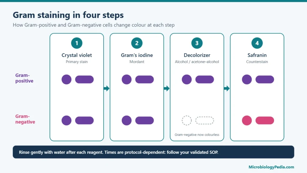
Gram-positive bacteria keep the crystal violet–iodine complex in their thick peptidoglycan wall and stay purple. Gram-negative bacteria lose it during decolorization and take up the pink safranin counterstain.
Escherichia coli Gram stain: positive or negative?
Escherichia coli (E. coli) is Gram-negative. On a Gram-stained smear it appears as pink to red, straight rods (bacilli) about 1–3 µm long, lying singly or in pairs, with no spores. The thin peptidoglycan layer and outer membrane do not retain crystal violet after decolorization, so the cells take up the safranin counterstain. The Gram stain cannot separate E. coli from other Enterobacterales such as Klebsiella or Enterobacter; lactose fermentation on MacConkey agar and biochemical tests (indole positive, citrate negative) are used for that.
Staphylococcus aureus Gram stain
Staphylococcus aureus (staph aureus) is Gram-positive. It appears as purple, round cocci about 1 µm in diameter arranged in irregular, grape-like clusters; pairs, short chains and single cells are also seen, especially in smears from broth or clinical material. Identification is confirmed by a positive catalase test and a positive coagulase test, and by yellow colonies with mannitol fermentation on mannitol salt agar.
Staphylococcus epidermidis Gram stain
Staphylococcus epidermidis (S. epidermidis) is Gram-positive and looks the same as S. aureus under the microscope: purple cocci in clusters. The two cannot be told apart by Gram stain. S. epidermidis is coagulase negative and does not ferment mannitol, and it is one of the organisms most often recovered from personnel monitoring in cleanrooms because it is part of normal skin flora.
Streptococcus pneumoniae Gram stain (pneumococcus)
Streptococcus pneumoniae (strep pneumo, pneumococcus) is Gram-positive. It appears as purple, lancet-shaped (elongated, slightly pointed) cocci in pairs, called diplococci, and sometimes in short chains. A clear halo around the cells may be visible where the capsule excludes the stain. It is catalase negative, alpha-hemolytic on blood agar and sensitive to optochin.
Haemophilus influenzae Gram stain
Haemophilus influenzae is Gram-negative. On Gram stain it appears as very small, pale pink coccobacilli that vary in shape (pleomorphic), from short rods to occasional filaments. Because the cells are small and stain faintly, they are easy to miss against the pink background of a clinical smear.
Listeria monocytogenes Gram stain
Listeria monocytogenes is Gram-positive. It appears as short, purple rods, single or in short chains, and may resemble coccobacilli or diphtheroids; older cultures can stain unevenly. It does not form spores, is catalase positive and shows tumbling motility at room temperature.
Klebsiella pneumoniae Gram stain
Klebsiella pneumoniae is Gram-negative. It appears as pink, short, plump rods, often surrounded by a clear zone because of its large polysaccharide capsule. It is non-motile and forms mucoid, lactose-fermenting colonies on MacConkey agar.
Neisseria meningitidis Gram stain
Neisseria meningitidis is Gram-negative. It appears as pink diplococci with flattened adjacent sides (kidney-bean or coffee-bean shape), and in clinical smears is often seen inside neutrophils. It is oxidase positive.
Enterococcus faecalis Gram stain
Enterococcus faecalis (E. faecalis) is Gram-positive. It appears as purple, oval cocci in pairs and short chains. It is catalase negative and grows and hydrolyzes esculin in the presence of bile (bile esculin positive).
Pseudomonas aeruginosa Gram stain
Pseudomonas aeruginosa is Gram-negative. It appears as pink, slender, straight or slightly curved rods, usually single. It is oxidase positive and does not ferment lactose.
Bacillus Gram stain: is Bacillus Gram-positive or Gram-negative?
Members of the genus Bacillus, such as Bacillus subtilis and Bacillus cereus, are Gram-positive: large purple rods, often in chains, that may show unstained oval areas where endospores have formed. Old cultures can lose the crystal violet and appear Gram-variable or pink. The word bacillus (plural bacilli) with a small b only describes a rod shape, so “Gram-negative bacilli” means rod-shaped Gram-negative bacteria such as E. coli, not the genus Bacillus.
Bacteria that do not Gram stain reliably
- Mycobacterium species: the waxy, mycolic-acid cell wall resists the stain; an acid-fast stain is used instead.
- Mycoplasma species: no cell wall, so they do not hold the stain.
- Old cultures of Gram-positive bacteria (for example Bacillus and Clostridium): damaged walls lose crystal violet and cells appear pink. Use cultures 18–24 hours old.
- Acinetobacter species: Gram-negative, but they resist decolorization and can appear Gram-positive even in a well-prepared smear.
- Over-decolorized smears: Gram-positive cells appear pink. Running a known Gram-positive and Gram-negative control on the same slide detects this.
What a Gram stain can and cannot tell
A Gram stain gives the Gram reaction, shape and arrangement within minutes, which narrows identification to a group. It does not identify the species: S. aureus and S. epidermidis, or E. coli and Klebsiella, look alike. Identification is completed with culture on selective and differential media, biochemical tests (catalase, coagulase, oxidase, IMViC) or an identification system.
Frequently Asked Questions (FAQs)
Is E. coli Gram-positive or Gram-negative?
E. coli is Gram-negative. It stains pink to red and appears as rods.
What does Staphylococcus aureus look like on a Gram stain?
Purple (Gram-positive) cocci in grape-like clusters, about 1 µm in diameter.
How is S. aureus distinguished from S. epidermidis?
Not by Gram stain; both are Gram-positive cocci in clusters. The coagulase test separates them: S. aureus is coagulase positive and S. epidermidis is coagulase negative.
What does Streptococcus pneumoniae look like on a Gram stain?
Purple, lancet-shaped diplococci (pairs), sometimes in short chains, often with a clear capsular halo.
Is Listeria Gram-positive?
Yes. Listeria monocytogenes is a short Gram-positive rod that does not form spores.
Why do Gram-positive bacteria sometimes look pink?
Usually because the culture is old, the smear was over-decolorized or the smear was too thick or overheated during fixing.
References
- Smith AC, Hussey MA. Gram Stain Protocols. American Society for Microbiology; 2005. asm.org/protocols/gram-stain-protocols
- Tripathi N, Sapra A. Gram Staining. In: StatPearls [Internet]. Treasure Island (FL): StatPearls Publishing; updated 2023. NCBI Bookshelf NBK562156
- Coico R. Gram staining. Current Protocols in Microbiology. 2005; Appendix 3C. doi:10.1002/9780471729259.mca03cs00
Related guides on MicrobiologyPedia
- Gram-positive vs Gram-negative bacteria: differences, cell wall and examples
- Gram staining: principle, procedure and results
- Gram-positive vs Gram-negative bacteria: differences and examples
- From Gram stain to culture: a practical reading sequence
- Microbial limit test (USP 61 and 62): tests for specified microorganisms
- Growth promotion test of culture media
For Gram-positive cocci, the catalase test is the usual next step to separate staphylococci (catalase-positive) from streptococci and enterococci (catalase-negative). See Catalase Test: Principle, Procedure and Interpretation of Results.

Noman Iftikhar is a microbiologist with an MPhil in Microbiology and professional experience in pharmaceutical microbiology. His work includes sterility testing, bacterial endotoxin testing, microbial limit testing, environmental monitoring, and microbiological quality control. Through MicrobiologyPedia, he shares practical, evidence-based guidance to help microbiology students and laboratory professionals understand testing procedures, laboratory practices, and quality standards.